
Figure 1. Voxel placement and sample spectra from dorsolateral prefrontal cortex (DLPFC).
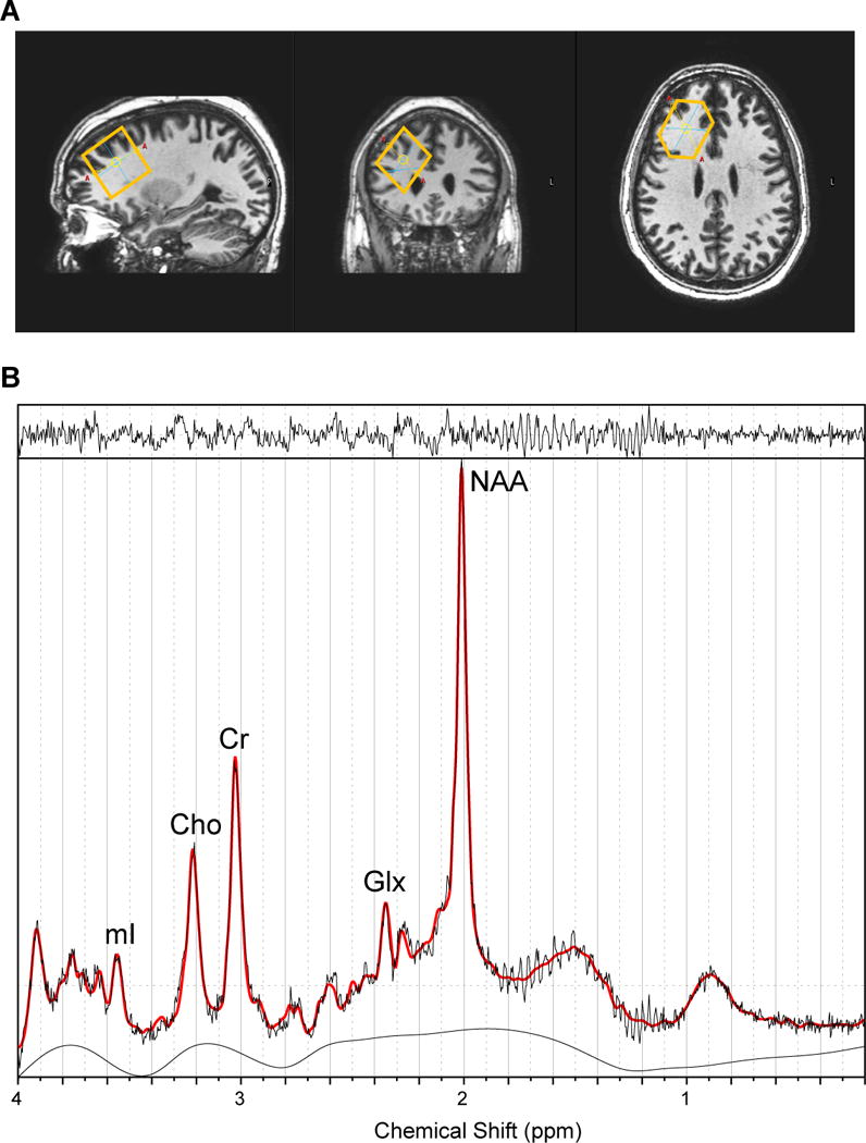
A. From left to right: sagittal view, coronal view, and axial view of the voxel.
B. A typical fitted spectrum (in red). Indicated are myo-inositol (mI), choline (Cho), creatine (Cr), Glx and total NAA. Above the spectrum the residual signal after fitting is displayed. The baseline is displayed below the spectrum.