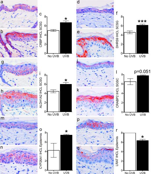
Figure 1. Acute UVB exposure localizes retinoid metabolism proteins to the upper epidermis.
SKH-1 mice were unexposed (a, d, g, j, m, p) or exposed (b, e, h, k, n, q) to 1 MED of UVB light and samples collected 48 hours later. Immunohistochemistry (IHC) was performed with antibodies against CRBP (a-c), DHRS9 (d-f), ALDH1A2 (g-i), CRABP2 (j-l), CYP26A1 (m-o), or LRAT (p-r). An IHC level (IHCL) was determined as the sum of intensity (0-4) plus percent of cells (0-4) for a maximum IHCL of 8. Bar = 10.1 uM. * p < 0.05, ** p < 0.01, *** p < 0.005.

