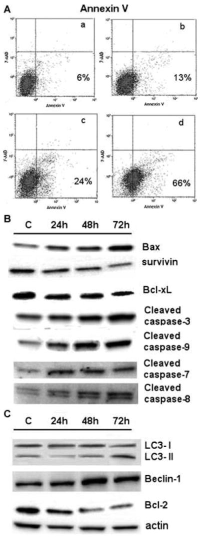
Fig. 3. Apoptosis and autophagy.

A. FACS measurement for apoptosis after culture of Hep3B cells for 48 h with 2.5 (a), 5 (b), 7.5 (c) or 10 (d) μM Regorafenib.
B and C. Western blotting of lysates from Hep3B cells treated with 7.5 μM Regorafenib for 24, 48 or 72 h and probed for apoptosis (B) and autophagy (C) markers.
C, controls (solvent alone, drug-untreated cells). The β-actin protein was used as loading control.
