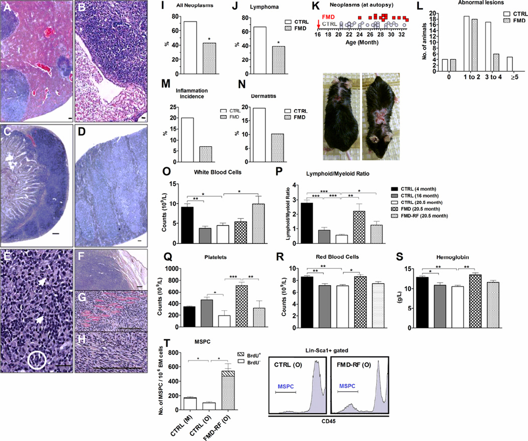
Figure 2. Periodic FMD cycle reduce and delay cancer, rejuvenate the hematopoietic system and induce mesenchymal stem/progenitor cells.
A) Hepatic lymphomatous nodules (bar= 400 microns). B) Lymphoma in the renal medulla (bar= 100 microns), C) in a mesenteric lymph node (bar= 100 microns) and D) in the spleen (bar= 100 microns). E) Hepatic lymphoma containing atypical cells with abnormal DNA (circle) and mitosis (arrows, bar= 100 microns). Subcutaneous fibrosarcoma in relationship to F) the epidermis and with invasion into G) the skeletal muscle tissue. H) Cytological details (bar= 100 microns). I) Autopsy-confirmed neoplasms. J) Lymphoma incidence. K) Neoplasms in relationship to the onset (arrow) of the FMD diet. L) Number of animals with 0 to more than 5 abnormal lesions determined at autopsy. M) Inflammatory incidence. N) Dermatitis incidence in %. Images show progression of dermatitis. O) – T) Complete blood counts. N= 7-12/group. O) White blood cells, P) Lymphoid: myeloid ratio. Q) Platelets, R) Red blood cells and S) Hemoglobin. Other CBC parameters are summarized in Table S3 and Figure S8. T) lin−Scal-1+CD45− mesenchymal stem/progenitor cells (MSPC) in bone marrow cells from control mature (M, 8–10 month), old (O, 20.5 month), and FMD mice 7 days after refeeding (FMD-RF; 20.5 month). N= 4-5/group. All data are expressed as the mean ± SEM.

