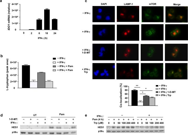
Figure 4. IDO-mediated tryptophan depletion suppresses mTOR lysosomal localization and HES1 protein expression.
(a) qPCR analysis of IDO1 mRNA in human primary macrophages treated with IFN-γ (100 U/ml) for 0-24 h (error bars, s.d.). Data are shown as means + SD of triplicate determinants and are normalized relative to GAPDH mRNA. (b) HPLC-MS measurement of intracellular L-tryptophan concentration in control or IFN-γ-primed human primary macrophages treated with or without Pam3CSK4 (10 ng/ml) for 4h (error bars, s.d.). Data are shown as means + SD of triplicate determinants. (c) Upper panels: immunofluorescence images of LAMP1 (red) and mTOR (green) co-staining in control macrophages (row 1), IFN-γ-primed macrophages (row 2), IFN-γ-primed macrophages pretreated for 30min with IDO inhibitor 1-D-MT (200 μM) (row 3), and IFN-γ-primed macrophages supplemented with tryptophan (Trp) (800 μM) (row 4). All cells were stimulated with Pam3CSK4 (10 ng/ml) for 4 h; nuclei were counter-stained with DAPI (blue). Lower panel: quantitation of co-localization between LAMP1 and mTOR (error bars, s.e.m.). Data are presented as mean + SEM of the percentage of co-localized cells from 800 cells counted in two independent experiments; overall p = 0.0008 by one way ANOVA followed by Bonferroni's multiple comparison post-test; *p<0.05, ** p<0.0001. (d, e) Immunoblot analysis of HES1 in control or IFN-γ-primed macrophages treated for 30min with 1-MT (200 μM) (d) or the indicated concentrations of tryptophan (Trp) (e), and then stimulated with Pam3CSK4 (10 ng/ml) for 4h; p38α serves as loading control. Data are representative of at least three (a, d, e) or two (b, c) independent experiments.

