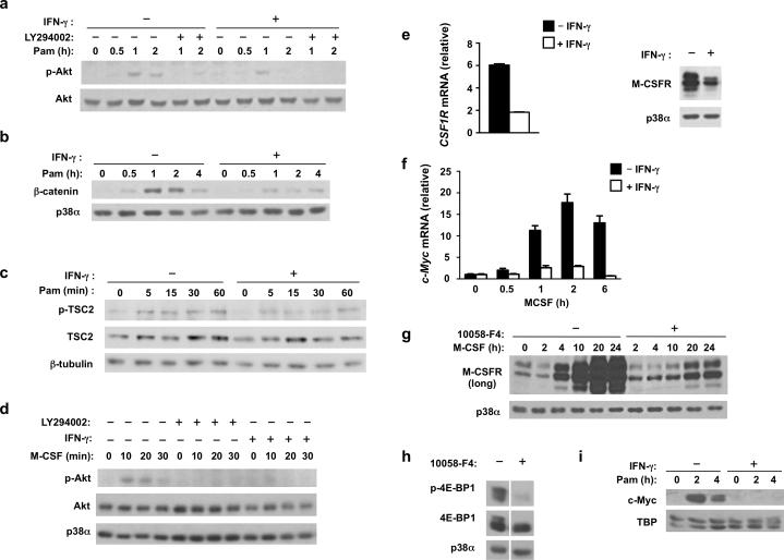
Figure 5. IFN-γ inhibits PI3K-Akt-TSC1/2 signaling and M-CSFR expression.
(a-c) Immunoblot analysis of whole cell lysates from control or IFN-γ-primed macrophages that were stimulated with Pam3CSK4 (10 ng/ml) for the indicated times. (d) Immunoblot analysis of phosphorylated (p-)Akt in control or IFN-γ-primed macrophages that were serum- and M-CSF-starved for 4 h, followed by pretreatment with vehicle control DMSO or LY294002 (10 μM) for 30 min, and then stimulated with M-CSF (100 ng/ml) for 0-30 min; Akt serves as loading control. (e) qPCR analysis (left panel) of CSF1R mRNA in control or IFN-γ-primed macrophages (error bars, s.d.). Data are shown as means + SD of triplicate determinants and are normalized relative to GAPDH mRNA. Immunoblot analysis (right panel) of M-CSFR in control or IFN-γ-primed macrophages; p38α serves as loading control. (f) qPCR analysis of MYC mRNA in monocytes cultured with M-CSF (20 ng/ml) with or without IFN-γ for indicated times (error bars, s.d.). Data are shown as means + SD of triplicate determinants and are normalized relative to GAPDH mRNA. (g) Immunoblot analysis of M-CSFR in human primary monocytes treated with vehicle control DMSO or Myc inhibitor 10058-F4 (60 μM) and then cultured with M-CSF (20 ng/ml) for indicated time points; p38α serves as loading control. (h) Immunoblot analysis of p-4E-BP1 in human primary monocytes treated with vehicle control DMSO or Myc inhibitor 10058-F4 (60 μM) for 30 min. (i) Immunoblot analysis of c-Myc in nuclear extracts of control or IFN-γ-primed macrophages that were stimulated with Pam3CSK4 (10 ng/ml) for 0-4h; TBP serves as loading control. Data are representative of at least three independent experiments (a-i).

