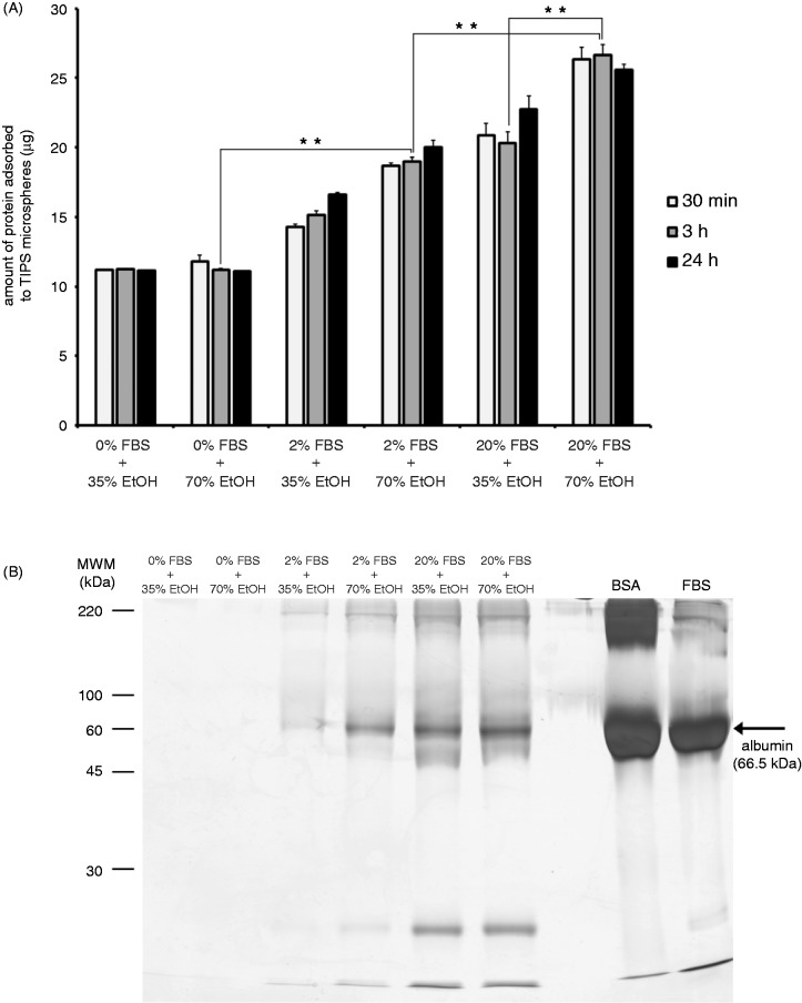
Figure 5.
The adsorption of serum proteins to PLGA microspheres is dependent on the extent of pre-wetting. PLGA microspheres were suspended in medium containing 0%, 2% or 20% FBS and treated with 35% (v/v) or 70% (v/v) ethanol (EtOH), for 30 min, 3 h and 24 h. The total amount of serum protein adsorbed to microspheres was measured after each incubation period (a). Serum and EtOH-treated microspheres were heated (95℃) in Laemmli buffer containing the reducing agent β-mercaptoethanol, proteins were separated using 15% acrylamide gels and stained using silver nitrate (b). Bovine serum albumin (BSA) and fetal bovine serum (FBS) were included as external controls. Data points represent the mean (n = 3 ± SEM) amount of total protein. *p ≤ 0.05 indicate differences between wetting conditions. Gel represents three individual experiments from three different sets of microsphere-protein adsorption experiments.

