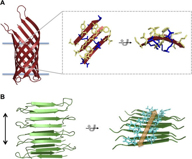
Fig 1. Illustration of protein structures.
(A) Cartoon depiction of the transmembrane domain of OmpA (PDB id 1QJP [3]) with the membrane indicated by blue horizontal lines. A zoomed-in section of β-sheet is shown in stick representation with hydrophobic side chains depicted in yellow and hydrophilic side chains depicted in blue. A rotated side view of the β-sheet section is shown on the right. (B) Cartoon depiction of the cross-β structure of the amyloid-forming peptide D23N-Aß1-40 (PDB id 2LNQ [4]). The fibril axis is indicated by a vertical black arrow. A rotated view of the structure is shown on the right (only one β-sheet is shown for clarity) to illustrate the proposed binding groove for the dye Thioflavin T. Side chains lining the groove are shown as cyan sticks and the binding groove is indicated by an orange arrow.

