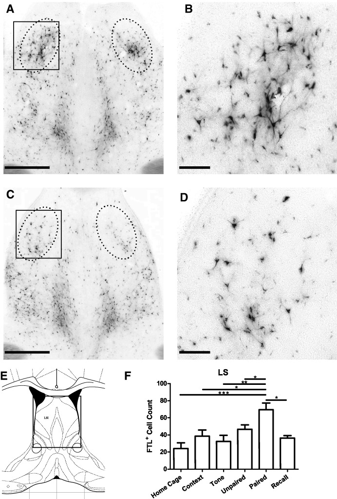
Figure 8.
Learning-specific FTL+ neurons in lateral septum (LSI) following auditory fear conditioning. (A,C) Bright-field images of LSI at bregma 0.3 mm from a Paired and Unpaired mouse, respectively. The solid box encloses the area shown in high power in B. Counted region is encompassed by dotted line. (B,D) High-power views of FTL+ neurons in Paired and Unpaired LSI, respectively. (E) Plate from Mouse Brain Atlas (Franklin and Paxinos 2008) highlighting the region shown in A and C. (F) FTL+ neuron counts of LSI region in each group of trained mice, shown as mean ± SEM. Significantly more FTL+ neurons were present in Paired mouse LSI region compared with controls. (*) P < 0.05, (**) P < 0.01, (***) P < 0.001. Scale bar, 500 µm (A, C), 100 µm (B, D).

