
FIGURE 2.
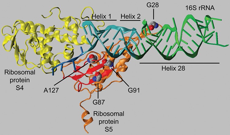
The position of the mutant amino acids of S5 (in orange) in relation to S4 (in yellow) and the central pseudoknot (helices 1 and 2; in cyan) and the neck helix (helix 28; in green) in 30S subunit (PDB ID 2AVY). The three amino acids altered in the S5 mutants described here (G87, G91, and A127) are shown in space-filling mode. The residues are located in a domain of S5 (in red) including a β sheet, composed of two β strands, and an adjoining strand. This domain is in close proximity to the central pseudoknot. The position of the spectinomycin-resistance mutant G28 is also shown in space-filling and is located near the region of h28 that is stacked on h2. The structure was modeled using VMD (Humphrey et al. 1996).