
Figure 2.
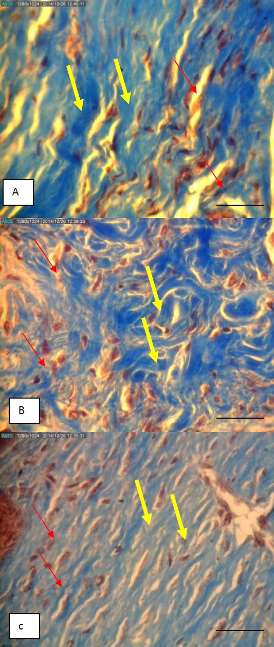
Microscopic pictures of derm skin of mice that showed thickness of collagen fiber and color density in the Malva sylvestris group (A), silver sulfadiazine group (B) and cold cream group (C) on day 10 (Trichrome masson staining, X400); yellow arrow: collagen fibers, red arrow: fibroblast cells; bar = 50 µm