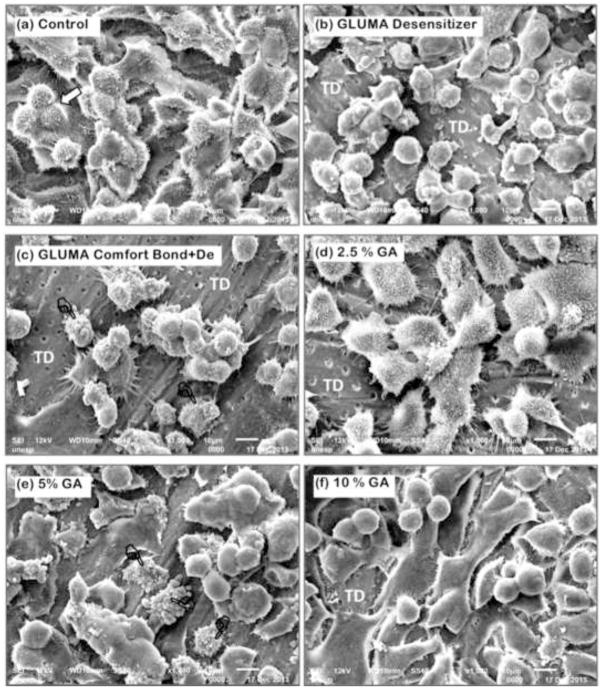
Figure 3.
General view of the odontoblast-like MDPC-23 cells seeded on dentin substrate. SEM, x1000. (a) Control: Cells with large cytoplasm covering most of the dentin surface. No dentinal tubules are visible. These cells have a number of processes which seem to attach them to the substrate. Note the occurrence of cell mitosis (white arrow). (b) Gluma desensitizer: A number of cells that remained adhered to dentin have round shapes and less cytoplasm fewer cytoplamatic projection, making them smaller than in the control group. (c) Gluma Bond: Reduced number of round-shaped MDPC-23 cells remained attached to dentin. Note that a large surface of tubular dentin (TD) with no cells is exposed. Cells with disrupted cytoplasm membrane can also be seen (pointers). (d) 2.5% GA: Cells with large cytoplasm, such as observed in control group, are attached to dentin. However, small surface of tubular dentin (TD) with no cell can be seen. (e) 5% GA: A number of MDPC-23 cells with variable morphology are observed. Note that a few cells show cytoplasm disruption (pointers). (f) 10% GA: Such as observed in (e), cells with different morphology are covering the dentin substrate. Most of these cells show only a few thin and short cytoplasm processes.

