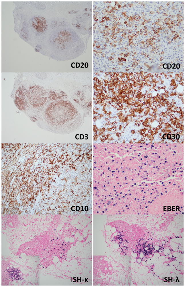
Figure 3.
Immunohistochemistry of case #1. The nodular aggregates of medium to large cells are CD20+. CD3 shows a background of smaller appearing cells. CD30 is positive in many immunoblasts. CD10 shows positive staining in stromal elements, but is also positive in small lymphocytes. EBER is positive in a large number of immunoblasts. In-situ hybridization reveals an overall polyclonal pattern. While some areas show lambda predominance, others show the opposite (small inlet images).

