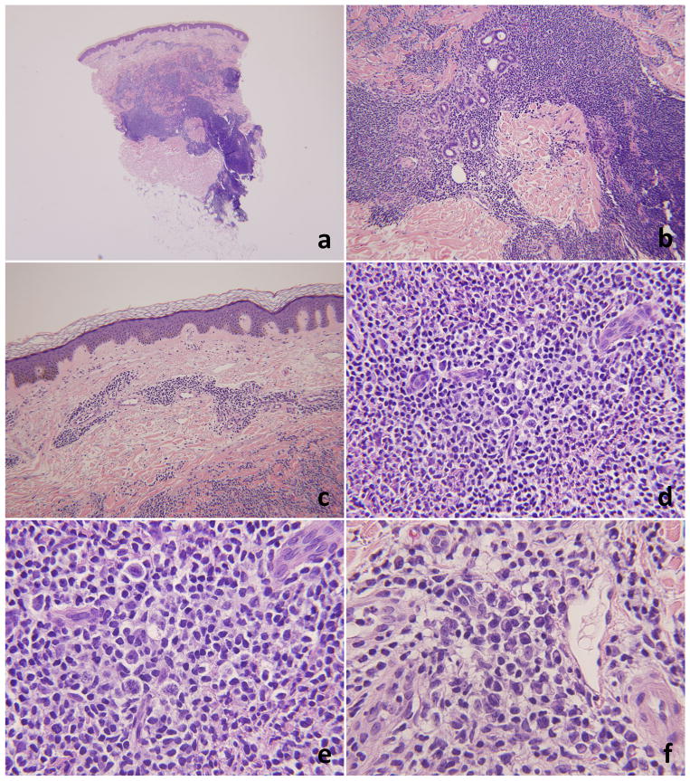
Figure 4.
Histopathology of case #2. 4a and 4b low and mid power views (20× and 100×). There is a nodular and diffuse lymphoid infiltrate in the superficial and deep dermis with focal extension into the subcutis. The infiltrate is sparing the epidermis (4c, 100×). 4d and 4e – high power views (200× and 400×, respectively). The infiltrate is heterogenous and includes a large population of immunoblasts. Admixed plasma cells are frequently seen (4f, 400×).

