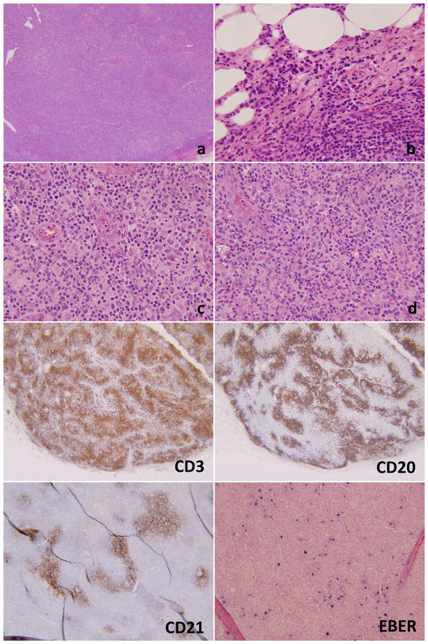
Figure 6.
Lymph node biopsy of case #2. There is effacement of the architecture at low power magnification (6a, 20×). Numerous plasma cells are seen at the periphery of the lymph node (6b, 200×). 6c and 6d (400×, each) show very prominent vasculature with high endothelial venules and a background very rich in eosinophils. The immunostains show a predominance of CD3+ T-cells as compared to the CD20+ B-cells. CD21 shows arborizing and disrupted follicular dendritic networks. EBER is positive in numerous lymphoid cells.

