Figure 1.
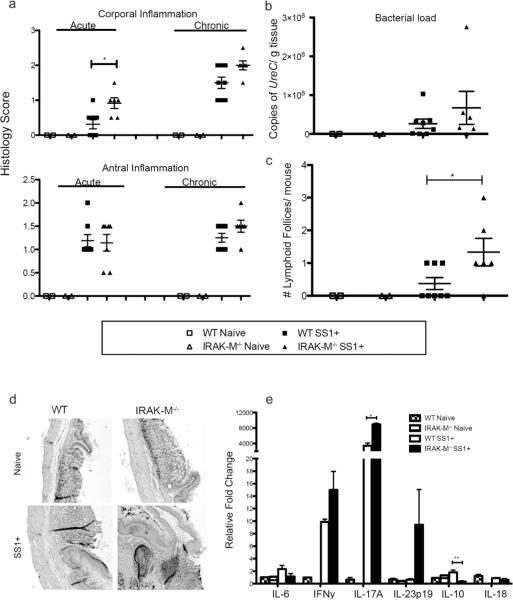
IRAK-M expression limits the development of H. pylori associated lymphoid follicles . WT and IRAK-M KO mice were infected with H. pylori for 16 weeks (n ≥ 6) (a) Acute and chronic inflammation were scored separately for the corpus and antrum on a scale of 0 – 3 (± SEM) (b) Bacterial load was determined by PCR quantification of ureC gene copy number per gram of stomach tissue (± SEM) (c) The number of lymphoid follicles present along the entire length of the gastric mucosa using histologic sections was determined (± SEM). (d) Representative H&E stained stomach sections demonstrating the primary location of lymphoid follicles (100X). (e) Cytokine expression was determined by semi-quantitative PCR using RNA isolated from gastric tissue (± SEM). * P<0.05, ** P<0.01.

