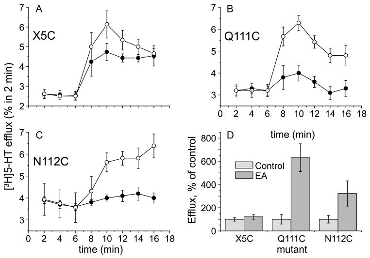
Figure 9.
Effect of MTSEA on [3H]5-HTefflux induced by extracellular 5-HT. Representative efflux experiments of X5C-SERT (panel A; n = 6–7), Q111C-SERT (panel B; n = 5–13, all experiments performed in triplicate) and N112C-SERT (panel C; n = 10–11) stably expressed in HEK293 cells. Cells were grown on glass coverslips and preloaded with tritiated serotonin for 20 min in the presence (open symbols) or absence of MTSEA (2.5 mM; filled symbols). After a subsequent wash step, coverslips were transferred to small superfusion chambers and superfused (for details, see ‘Materials and methods’). The experiment was started after a 45 min washout period at t = 0 with the collection of superfusate in 2-min fractions. 5-HT (10 μM) was added to the superfusion buffer after 6 min. The collected radioactivity was counted and is expressed as a fractional efflux rate, i.e., as a percentage per 2 min of the cellular [3−H]5-HT content at that very time point. Baseline efflux was adjusted for more accurate comparison between control and MTSEA-treated cells. Panel D summarizes the integrated effects of 5-HT treatment in the presence or absence of MTSEA (as indicated) with subtraction of baseline efflux levels.

