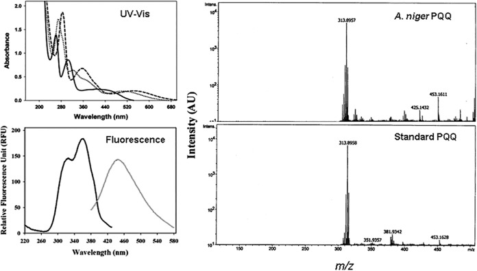
FIG 5.
Identification of PQQ in A. niger spent medium. (Left) The UV-Vis spectra (solid line, pH 1.0; dotted line, pH 7.0; dashed line, pH 12.0) and the fluorescence spectra at pH 7.0 (black line, emission λ at 448 nm; gray line, excitation λ at 323 nm) of the isolated pink compound are shown. (Right) The mass spectrum of the pink compound is compared with that of standard PQQ. AU, relative abundance units.

