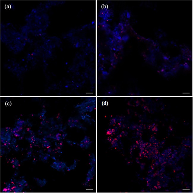
FIG 4.
FISH images of the inoculum (a) and of the enrichment culture after 9 (b), 16 (c), and 20 (d) months. The fluorescence micrographs were taken after hybridization with the NC10 bacterium-specific probe S-*-DBACT-1027-a-A-18 (Cy3, red) and the general DNA stain DAPI (blue). The “Ca. Methylomirabilis oxyfera”-like bacteria appear magenta due to a mixture of the two fluorescence signals (red and blue). Bar, 10 μm.

