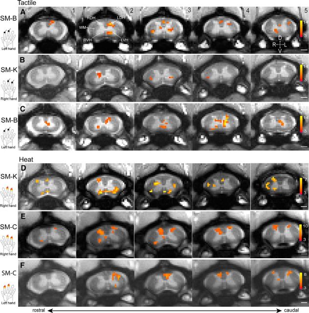
Figure 2.
Representative fMRI activations to tactile and nociceptive heat stimulation of two distal finger pads in three monkeys (SM-K, SM-C, and SM-B). A, Single-run fMRI activations to tactile stimulation of two distal finger pads on right hands. B, C, Multirun average fMRI activations to tactile stimulation of two distal finger pads on right (B) and left (C) hands. D–F, Multirun average fMRI activations to 47.5°C nociceptive heat stimulation of two distal finger pads on right (D, E) and left (F) hands. Hand inserts show the locations of stimulation. All activation maps are thresholded at p < 0.05 for multirun and p < 0.01 for single run with FDR corrected; see color scale bar on image 5 for the t value range. Images 1–5, From caudal to rostral. Scale bars, 1 mm. D, Dorsal; V, ventral; L, left; R, right.

