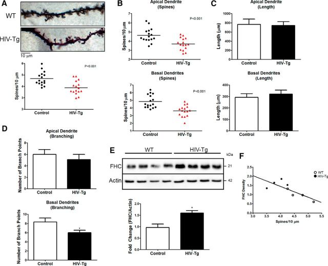
Figure 1.
Dendritic spine density and branching are reduced in the PFC of the HIV Tg rat and correlates with FHC changes. A, Overall dendritic spine density of layer II/III pyramidal neurons in the medial PFC of HIV Tg rats is significantly reduced compared with F344 WT controls. Data shown in A–D come from four animals per group; four neurons were analyzed for each brain (16 neurons per group). B, Both apical and basal dendritic spine density is decreased by a similar magnitude in HIV Tg rats. C, Apical and basal dendrite length is unaltered in HIV Tg rats compared with WT controls. D, Analysis of branching in apical and basal dendrites revealed a significant decrease in the number of branch points in basal dendrites of HIV Tg animals but not apical dendrites. E, Frontal cortex lysates of HIV Tg rats used for Neurolucida analysis are shown to have increased FHC levels compared with control animals (n = 4 per group). F, Plotting each animal's FHC protein levels to their overall spine density revealed a significant inverse correlation, indicating a negative relationship between PFC FHC protein levels and spine density (n = 8; Pearson's r = −0.7815, p = 0.0220). *p < .0.05; red symbols denote p < 0.001.

