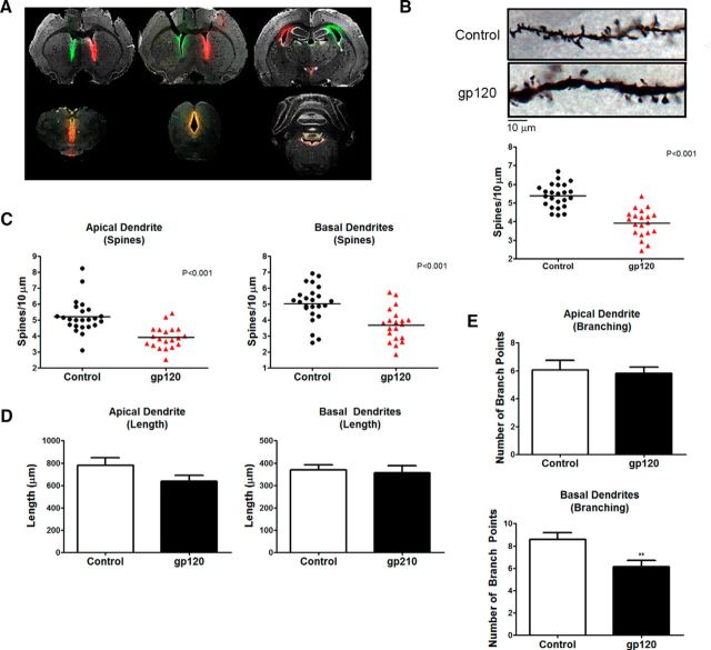
Figure 2.
Rats treated with gp120IIIB for 7 d exhibited decreases in dendritic spine density and branching. A, Coronal sections through the forebrain, midbrain, and brainstem show the results of bilateral intracerebroventricular infusion of fluorescent tracers into the rat brain using stainless-steel cannulas, as described in Materials and Methods. Evidence of tracer diffusion was observed throughout lateral, third, and fourth ventricles. B, Overall dendritic spine density of layer II/III pyramidal neurons in the medial PFC of gp120-treated SD rats is significantly reduced compared with vehicle-treated controls. Data shown in B–E come from four neurons per brain (i.e., control, n = 24; gp120, n = 20). C, In gp120-treated rats, apical and basal dendritic spine density is significantly reduced by a similar magnitude. D, The number of basal branch points, but not apical branch points, is significantly less in gp120-treated rats compared with control animals. E, Both apical and basal dendrite length are not different between control and gp120-treated rats. **p < 0.01; red symbols denote p < 0.001.

