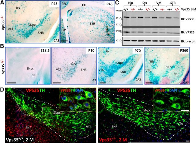
Figure 1.
Expression of VPS35 in DA neurons. A, Detection of the β-gal activity (blue color) in VPS35+/− SNpc, VTA, red nucleus (RN), STR, and hippocampus CA3 at P45. cc, Corpus callosum. The β-gal activity is used as a reporter for VPS35's expression, because the LacZ gene is inserted in the intron of the VPS35 gene in VPS35+/− mouse. Scale bar, 200 μm. B, Detection of the β-gal activity in VPS35+/− VM from different aged mice (E18.5, P10, P70, and P360). In young adult brain (e.g., P70), β-gal activity reached the peak level in VTA and SNpc, which remained positive in 1-year-old or older mice. Scale bars, 200 μm. C, Western blot analysis of retromer complex VPS35 (∼90 kDa) and VPS26 (∼36 kDa) protein levels in homogenates of Hip, Ctx, VM, and STR of 8-month-old VPS35+/+ and VPS35+/− brains. Note that ∼50% reductions in both VPS35 and VPS26 protein levels were detected in homogenates from VPS35+/− brain samples. D, Coimmunofluorescence labeling of TH (green) and VPS35 (red) in the SNpc brain sections from 2-month-old (2 m) VPS35+/+ and VPS35+/− mice. The VPS35 immunoreactivity is reduced in TH+ neurons from VPS35+/− SNpc (right). Insets marked by yellow dashed rectangles are amplified images. Scale bars: 80 μm; inset, 10 μm. PFC, prefrontal cortex; IB, immunoblot.

