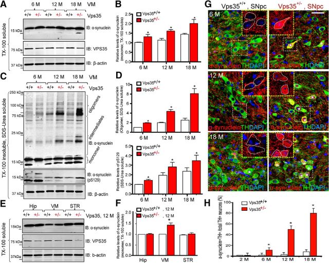
Figure 4.
Accumulation of α-synuclein proteins in VPS35+/− SNpc-DA neurons. A–D, Western blot analysis of soluble and insoluble α-synuclein levels from VM of various aged (6-, 12-, and 18-month-old) VPS35+/+ and VPS35+/− mice. TX-100 soluble (A, B) and insoluble (SDS/Urea soluble; C, D) fractions were isolated and analyzed via Western blotting using indicated antibodies. VPS35 was used to confirm VPS35+/− mice, and β-actin was used as a loading control. Data were quantified and presented in B and D as mean ± SEM; n = 3; *p < 0.01. E, F, Western blot analysis of soluble homogenates of α-synuclein levels from various brain regions (VM, STR, and Hip) of 12-month-old VPS35+/+ and VPS35+/− mice. Data in E were quantified and presented in F as mean ± SEM; n = 3; *p < 0.01. G, H, Coimmunofluorescence staining analyses of α-synuclein and TH in SNpcs from 6-, 12-, and 18-month-old VPS35+/+ and VPS35+/− mice. Coronal midbrain sections from VPS35+/+ and VPS35+/− mice at indicated ages were stained with α-synuclein and TH antibodies. Asterisk, Indicates the cell with somatic accumulation of α-synuclein. Scale bar, 10 μm. Quantitative analysis of the DA (TH+) neurons with somatic accumulation of α-synuclein in VMs at indicated ages was presented in H (mean ± SEM; n = 3; *p < 0.01). IB, immunoblot.

