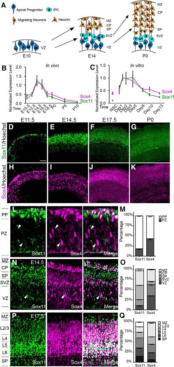
Figure 1.

Sox11 and Sox4 are dynamically expressed during corticogenesis in overlapping and discrete cellular populations. A, Schematic of corticogenesis with apical progenitors in the VZ (bright blue), IPCs in the SVZ (lighter blue), and neurons in the PP, SP, MZ, and CP (red). B, C, Profiles of Sox11 (green) and Sox4 (magenta) levels in cortical development (B) and in cortical cultures (C) obtained using qRT-PCR and normalized to U6 levels. D–K, Expression of Sox11 (D–G) and Sox4 (H–K) in mouse cerebral cortex at E11.5 (D, H), E14.5 (E, I), E17.5 (F, J), and P0 (G, K) with white lines indicating the position of the ventricle and pial surface (D, H) or ventricle, lower CP, and pial surface (E–K). L–Q, Expression (L, N,P) and quantification of distribution (M, O, Q) of Sox11 (green) and Sox4 (magenta) at E11.5 (L, M), E14.5 (N, O) and E17.5 (P, Q). Arrowheads indicate Sox4+Sox11−− cells in the apical cerebral wall (VZ and SVZ) and white cells reveal overlap of Sox11 and Sox4. Scale bars: D, E, H, I, 64 μm; F, G, J, K, 96 μm; L, 24 μm; N, P, 40 μm.
