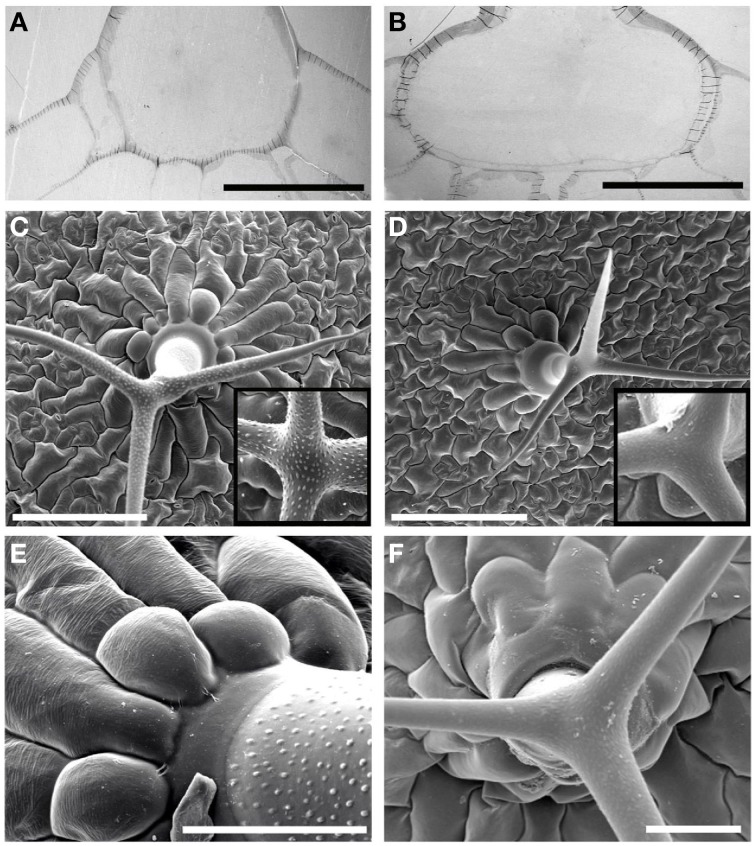
Figure 6.
Ultrastructural organization of the trichomes. Transverse ultra-thin sections of the trichomes in the wild type (A), and rwa2-3 (B). Scale bars equal 50 μm. Lines across the cell wall are artifacts due to wrinkling of the ultra-thin sections. Scanning electron micrographic images of trichomes in the wild type (C) and the rwa2-3 (D). Scale bars equal 200 μm. Inserts (100 × 100 μm) represent close-up images of trichome branches. Close-up images of the trichome base in the wild type (E) and rwa2-3 (F). Scale bars equal 50 μm. All images were acquired by electron microscopy.

