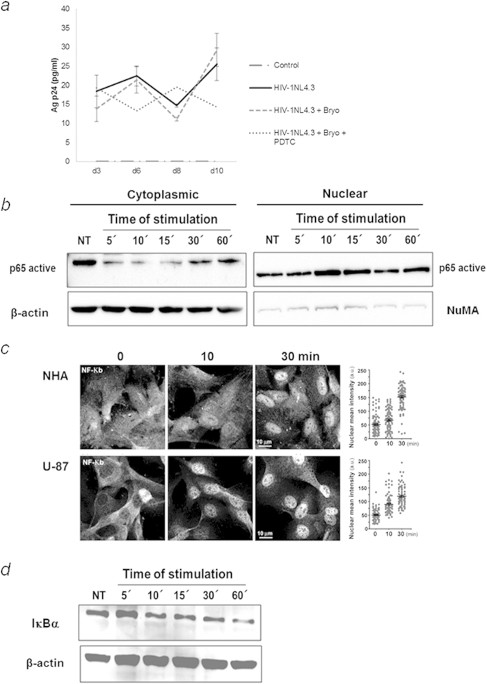
Figure 3. Bryostatin induces viral reactivation in a NF-κB-dependent manner.
(a) The p24 levels were monitored 2 and 5 dpi after treatment with bryostatin alone or in combination with PDTC (10 μM) in NHA supernatants. The mean values (mean ± S.D.) of three independent experiments are shown. (b) NHA cells were treated with 100 ng/ml bryostatin for the indicated times. Nuclear translocation of NF-κB upon treatment was measured using Western blotting. Antibodies directed against β-actin and NuMA were used as protein loading controls for the cytoplasmic and nuclear fractions, respectively. (c) Confocal immunofluorescent analysis of NHA and U-87 treated with or without bryostatin (left) using Phospho-NF-κB p65 (Ser536) Rabbit mAb (green). Nuclei were labeled with TOPRO. The nuclear:cytoplasmic ratios of immunostaining were measured at the single-cell level by quantifying the NF-κB intensities inside and outside of the nucleus (blue TROPO). Data for 500 single-cell measurements are shown for cells treated with or without NHA and bryostatin (100 ng/ml) for the indicated times (100 ng/ml) (right panel). Scale bar, 10 μm. (d) Bryostatin induces IκBα degradation. U-87 cells were cultured with bryostatin for the indicated time points (0–60 min). The cell lysates were blotted with antibodies specific for IκBα. Western blot data are representative of 3 independent experiments.

