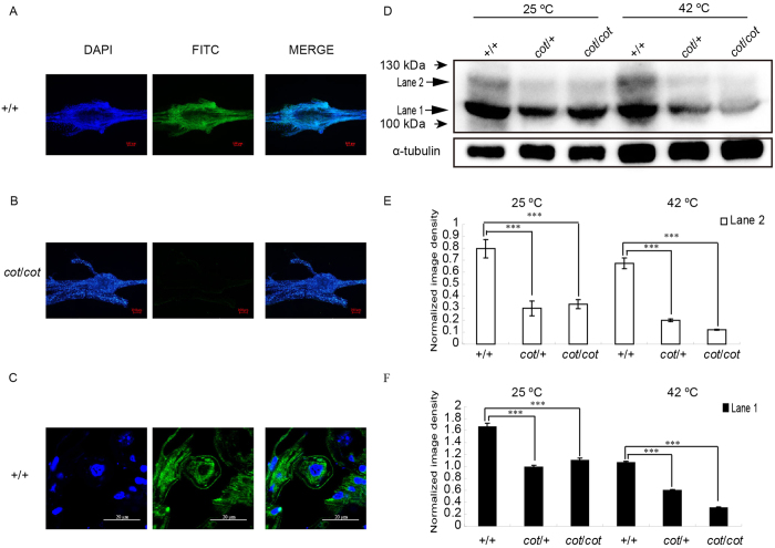
Figure 3. Expression and localization of Bmsei in the ganglia with immunofluorescence and immunoblotting.
Immunofluorescent signal for Bmsei in whole ganglia of +/+ larvae on day 5 of fifth instar (A), with a weak signal in cot/cot larvae (B). (C) Immunofluorescence in paraffin sections of +/+ ganglia on day 5 of fifth instar. Bmsei was localized in the cell membranes and cytoplasm. Green, positive signal; blue, DAPI staining of the nuclei. (D) Immunoblotting of Bmsei in the total proteins from new larvae of the three genotypes under normal conditions (25 °C) and after temperature stimulation (42 °C, 5 min). Bmsei appeared as two bands (lanes 1 and 2) in the WT. Both bands were significantly less intense (lower abundance) in the samples from cot/cot and cot/+ individuals than in samples from the WT (E and F). ***P < 0.001. Error bars depict s.e.m.

