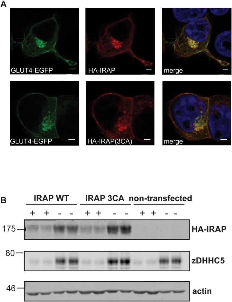
Figure 4. Intracellular localisation of wild-type and cysteine mutant IRAP.
A HEK293T cells were co-transfected with HA-tagged IRAP constructs together with GLUT4-EGFP. After 24 h, cells were fixed in 4% formaldehyde and subsequently permeabilised with Triton X-100. Cells were then incubated in HA antibody and subsequently in secondary antibody conjugated to Alexa Fluor 543. Cells were mounted in ProLong Gold Antifade mounting medium containing DAPI. Whole cell image stacks were acquired by confocal microscopy. Representative sections of a typical cell are shown. Separate channels displaying GLUT4-EGFP (green) and HA-tagged IRAP WT or 3CA mutant (red) and a merge of both channels including DAPI staining are shown. Scale bar = 5 μm. B Cells were transfected for 24 h with plasmids encoding HA-tagged IRAP wild type or cysteine to alanine mutant (3CA) and were incubated in trypsin-EDTA (0.05%) (+) or in standard culture media (–) for 30 minutes. Cell lysates were subjected to SDS-PAGE followed by immunoblotting with the indicated antibodies. Anti-HA was used for detection of HA-IRAP. Samples were loaded in duplicate and representative immunoblots are shown, with the position of molecular weight markers indicated on the left hand side of blots.

