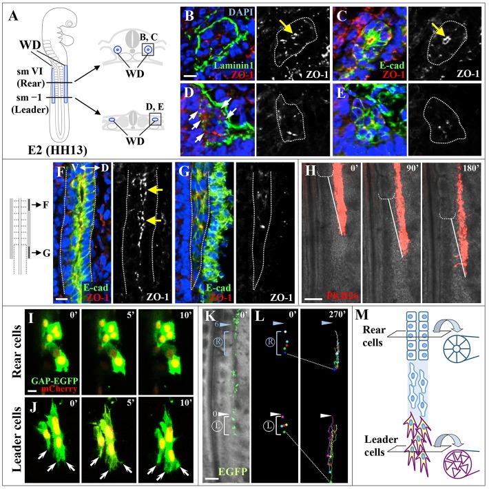
Fig. 1.
Cell morphology of leader and rear cells in the forming WD. (A) Drawings of whole-mount and transverse sections (in B-E) of an E2/HH13 chicken embryo. Cells located in the rear part [somite level (sm) V to VII] and the leader part (sm –2 to I) of the WD were termed rear cells and leader cells, respectively. (B-E) Transverse sections were stained with antibodies for laminin 1 and ZO-1 (B,D) or E-cadherin and ZO-1 (C,E). Yellow arrows (B,C) indicate apically localized ZO-1 in rear cells. White arrows (D) indicate sites where signals for laminin 1 are absent. Nuclei were stained with DAPI. (F,G) Sagittal sections of rear cells (F) and leader cells (G) were immunostained for E-cadherin and ZO-1. Yellow arrows (F) indicate lumens with apical ZO-1 signals. The E-cadherin-positive cell population seen dorsal to the WD is surface ectoderm. (H) Selected frames of an in vivo time-lapse movie (supplementary material Movie 1) showing the elongation of PKH26-labeled WD (red). White dotted brackets denote a newly formed somitic boundary. White solid lines indicate the interval between the white bracket and a tip of elongating WD. Note that the white lines in each panel are constant in length. (I,J) Selected frames from in vivo time-lapse movies (supplementary material Movies 2 and 3) showing magnified rear cells (I) and leader cells (J). Lamellipodia and filopodia were observed on leader cells (white arrows). (K,L) Migratory tracks of rear cells and leader cells are bracketed by blue and white lines, respectively. The light blue and white arrowheads indicate the sixth and newly formed somitic boundaries, respectively. (M) Diagram illustrating differential cell morphology in the elongating WD of the E2/HH13 chicken embryo. Leader cells are mesenchymal in shape and highly motile, whereas rear cells are static and constitute an epithelial tubule. Time is in minutes. Scale bars: 20 μm in B,F,I; 100 μm in H,K.

