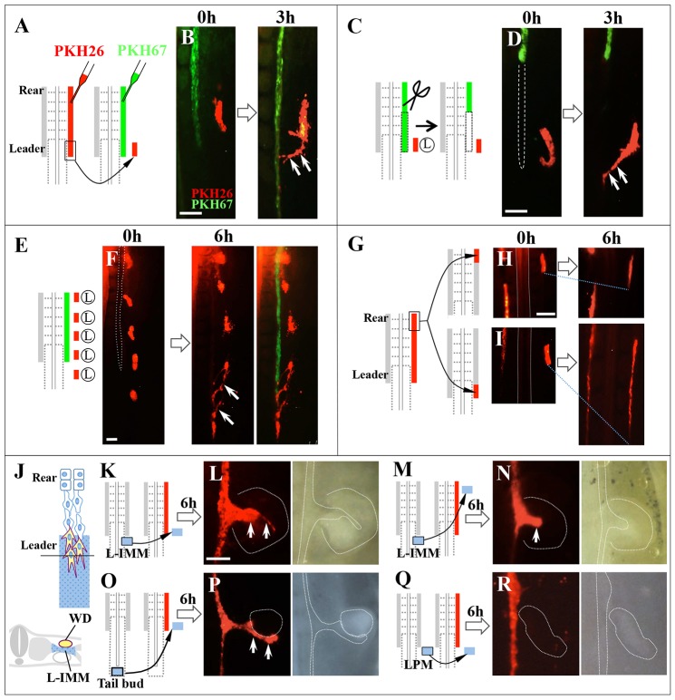
Fig. 2.
WD cells are attracted to IMM that neighbors leader cells. (A) PKH26-labeled leader cells (red) were transplanted into a region lateral to PKH67-labeled leader cells (green) of an E2/HH13 host embryo. (B) Donor cells (red) moved toward the host WD by 3 h after transplantation (arrows). (C) Leader cells (red) were grafted (similarly to B) into a host embryo from which leader cells had been removed. (D) The grafted leader cells moved mediocaudally (arrows), even if WD was not present. (E) Leader cells from five donor embryos (red) were transplanted into five different positions along the AP axis in the lateral plate mesoderm (LPM). (F) The posteriorly placed cells moved toward the leader area of host WD (arrows). (G-I) Rear cells of donor WD (red) were transplanted into the leader region (I) or rear region (H) of host embryos. (I) The transplant resumed marked elongation in the leader region. (J) The IMM adjacent to the leader cells was termed ‘L-intermediate mesoderm’ (L-IMM) (blue). (K,L) A piece of L-IMM was transplanted into LPM located medial to the leader cells. Arrows (L) indicate an ectopic branch from the host WD (red). (M,N) L-IMM also caused ectopic branch formation from the rear WD when relocated to this region (arrow). (O,P) A piece of tail bud was transplanted into LPM, resulting in the formation of an ectopic branch (arrows). (Q,R) LPM transplantation as a negative control. Scale bars: 100 μm.

