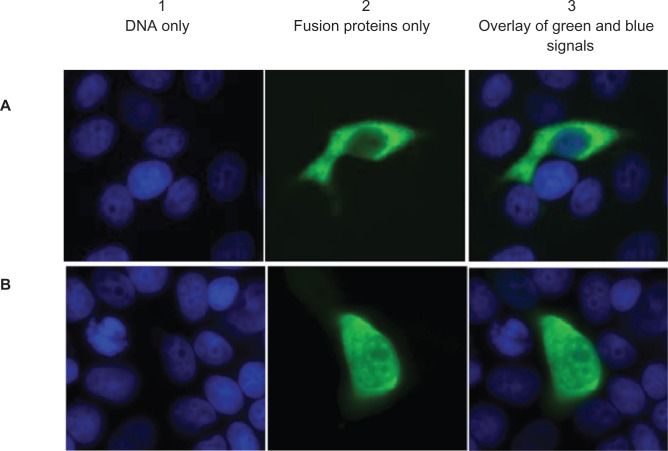
Figure 5.
Cellular localization of A) MLK4α and B) MLK4β c-myc fusion proteins. COS-1 cells were transfected with pCMV-MLK4α and pCMV-MLK4β constructs, and the c-myc-tag was stained with mouse α-c-myc monoclonal antibody (green). DNA is stained blue. (A1 and B1) DNA only; (A2 and B2) fusion proteins only; (A3 and B3) overlay of green and blue signals.

