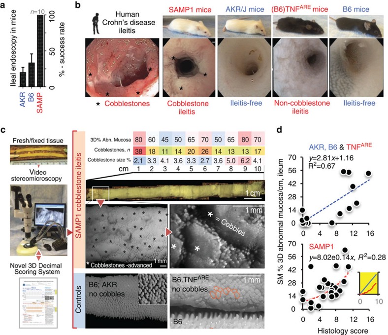
Figure 1. Stereomicroscopy allows the rapid topographical analysis of the entire intestinal mucosa in mice.
(a) Although small intestinal endoscopy is possible in mice, complications in healthy control mice due to tissue friability make endoscopy suboptimal/biased for routine murine GI examination. (b) Endoscopic images of terminal ileum from humans, ileitis-prone SAMP1 and TNFARE mice and ileitis-free genetic controls AKR/J and B6. Notice the similarity of obstructive cobblestone ileitis for humans and SAMP mice, and distinct endoscopic pattern in TNFARE. (c) To increase our understanding of intestinal diseases, we developed a 3D-SM Assessment and Pattern Profiling protocol (3D-SMAPgut) for fresh and fixed postmortem specimens to evaluate the mucosal architecture of both small and large intestine. The system characterizes abnormal/normal mucosa, cm by cm, using a reference catalogue of SM lesions, validated parameters (see Methods and Supplementary Figs 8–10). SM allowed the discovery of previously unknown ‘cobblestone' lesions in animals/mice. (d) Correlation between ileal SM (3D% of abnormal mucosa, 3D%-AbMuc) and traditional histological inflammation scores in mice. Notice histological scores plateau at 12 in SAMP (curved exponential fitting) compared with AKR and B6 (linear fit; higher R2). Inset line plot, simulation of SM 3D%AbMuc represented mathematically as circles whose cobblestone areas (area=πr2, red curved line) grow exponentially as their diameter increases. Radius, surrogate for histological score on intestine's longitudinal axis, increases as cobblestones grow (straight line, y=r). Lower R2 indicates larger variability between SM and histology in SAMP. SM-guided dissection of lesions decreases variability and increases biological/statistical study power.

