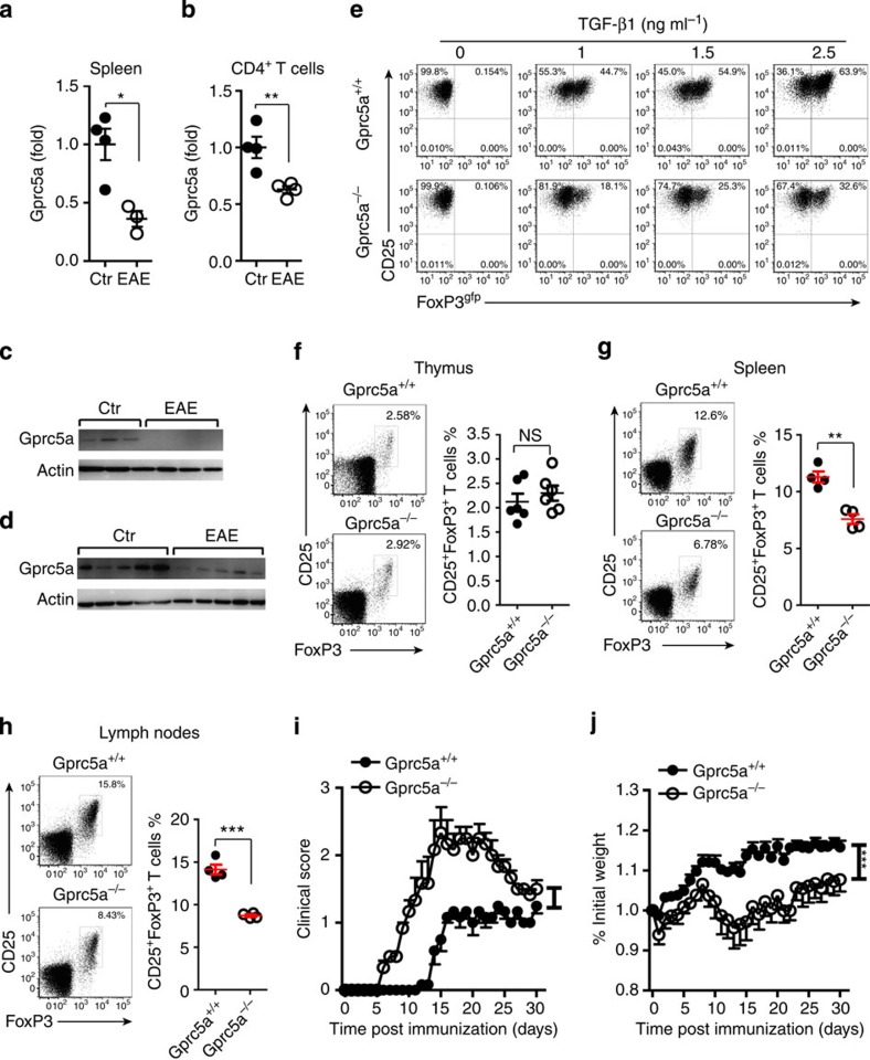
Figure 6. Gprc5a deficiency decreases pTreg-cell differentiation and promotes autoimmune inflammation.
(a,b) Splenocytes were prepared from unimmunized control mice (Ctr) or EAE mice 10 days after immunization (n=3–4). qPCR analysis of Gprc5a expression in total splenocytes or in sorted CD4+ T cells. (c,d) Western blot analysis of Gprc5a in splenocytes and CNS infiltrating cells derived from healthy controls (Ctr) or EAE mice. (e) Naive T cells were sorted from Gprc5a+/+ and Gprc5a−/− mice. Flow cytometry of polarized iTreg cells in the presence of different concentrations of TGF-β1. (f) Flow cytometric analysis of Treg cells in thymus from 6-week-old Gprc5a+/+ and Gprc5a−/− mice (gated on CD4+ T cells). (g,h) Flow cytometric analysis of Treg cells in the inflamed spleen and lymph nodes Gprc5a+/+ and Gprc5a−/− mice 14 days after the induction of EAE (gated on CD4+ T cells). Numbers adjacent to outlined areas indicate per cent cells in each. (i,j) Clinical scores and weight loss (mean±s.e.m.) of Gprc5a+/+ or Gprc5a−/− mice after the induction of EAE were assessed every day (n=7). *P<0.05, **P<0.01, ***P<0.001, NS, not significant, two-tailed Student's t-test for a,b,f,g and h; one-way analysis of variance for i,j. Data are representative of at least two independent experiments (mean±s.e.m.).

