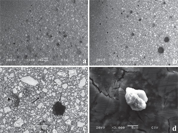
Figure 1. Blocks (4x4x1 mm) of (a) conventional FX-II, (b) FX-II 3% (w/w) TiO2, and (c) FX-II 5% (w/w) TiO2. Samples were gently polished and finished with #400, #1,000, and #1,500 waterproof abrasive paper and ultrasonically cleaned. Topographically, there are no differences between specimens. Nevertheless, hybrid particles are observed, microparticles (1c, black circle and arrow) are uniformly lay between (matrix) macroparticles, and such particles seem to be grouped of TiO2 nanoparticles due to their angular and semispherical shape confirmed by the 1d micrograph and EDS of this area, the zone exhibits higher concentration of titanium (a%=0.36%).

