Fig. 3.
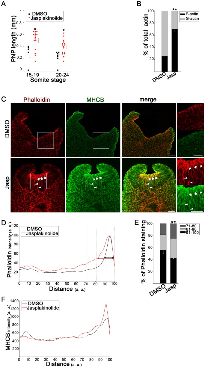
Stabilization of F-actin delays spinal neural tube closure. (A) PNP length is significantly increased at the 15–19 and 20–24 somite stage after 18–20 h culture in 10 nM jasplakinolide (Jasp), compared with DMSO controls (*P<0.05). (B) Biochemical fractionation shows proportionately increased F-actin and reduced G-actin in Jasp-treated embryos relative to DMSO controls (**P<0.001). (C) Immunohistochemistry (phalloidin, red; anti-MHCB, green) reveals actomyosin accumulation at the apical neuroepithelial surface (asterisks) and on some lateral cell surfaces (arrowheads) after Jasp treatment (embryos have 20–21 somites). Right, enlargement of the boxed areas. Scale bars: 30 µm. (D–F) Intensity profile scans along the neuroepithelial basal-to-apical axis. Jasp-treated embryos show an extension of phalloidin staining (intensity normalized to 100%) towards the basal surface (arrows in D), which is confirmed by quantification in the most apical 30% of the neuroepithelium (E; **P<0.001). MHCB staining intensity (non-normalized) is greater apically in Jasp-treated embryos than in DMSO controls (F). Significance values were calculated with a Student's t-test, compared with DMSO control.

