Fig. 4.
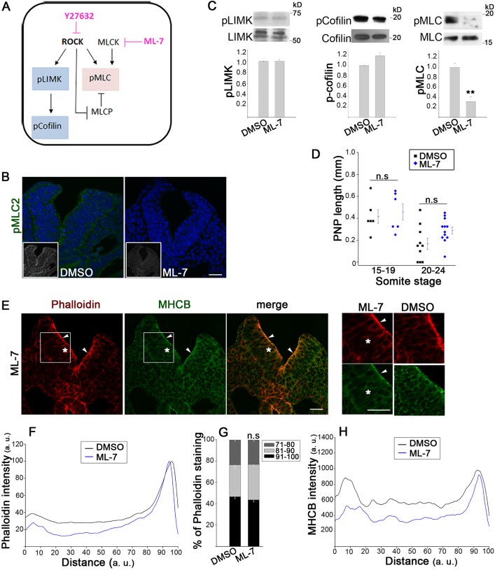
Spinal neurulation proceeds despite inhibition of myosin II contractility. (A) Downstream effectors of ROCK and inhibitory action of Y27632 and ML-7. (B) Immunohistochemistry shows absence of pMLC (green; nuclei, DAPI) from apical neuroepithelium of ML-7-treated embryos, compared with DMSO controls (embryos have 18–19 somites). Grayscale insets, pMLC staining only. Scale bar: 30 µm. (C) Western blots for pLIMK and LIMK, p-cofilin and cofilin, and pMLC and MLC. Embryos cultured for 5–6 h in ML-7 have significantly reduced the amount of pMLC compared with that in DMSO controls, whereas pLIMK and p-cofilin are unaffected (n=3, **P<0.001). (D) Culture for 5–6 h in 50 µM ML-7 does not significantly affect closure compared with DMSO at the 15–19 somite stage (n.s., P>0.05). (E) Immunohistochemistry (phalloidin, red; anti-MHCB, green) of ML-7-treated embryos shows apical actomyosin closely resembling DMSO controls (arrowheads; see insets on right), whereas actomyosin is reduced more basally (asterisks). Embryos have 18 somites. Scale bars: 30 µm. (F–H) Intensity profile scans show unchanged apical phalloidin staining after ML-7 treatment (F), as confirmed by quantification (G; n.s, P>0.05 versus DMSO). The more-basal phalloidin (F) and MHCB (H; non-normalized) staining is reduced by ML-7. Significance values were calculated with a Student's t-test, compared with DMSO control.

