Fig. 5.
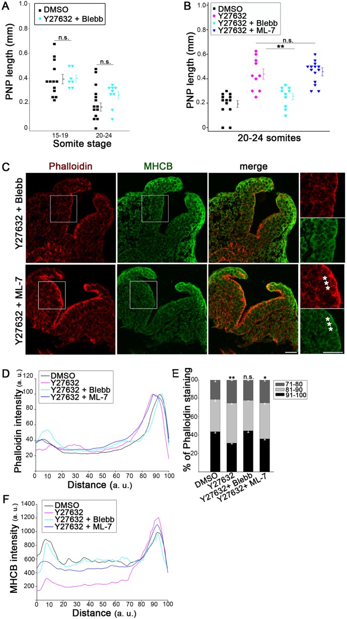
Prevention of Y27632-related closure defects by Blebbistatin but not ML-7. (A,B) The delayed PNP closure seen after culture in 5 µM Y27632 (B) is rescued by co-exposure to 50 µM Blebbistatin (A,B; **P<0.001) but not by ML-7 (B; n.s. P>0.05). Cultures were for 5–6 h (A) or 15–18 h (B), ending at the somite stages indicated. (C) Immunohistochemistry (phalloidin, red; anti-MHCB, green) reveals a normal actomyosin distribution in embryos co-exposed to Y27632+Blebbistatin (upper panels; compare with DMSO in Fig. 2A and Fig. 3C). In contrast, embryos co-exposed to Y27632+ML-7 (lower panels) show actomyosin accumulation apically (asterisks), as in those treated with Y27632 alone (see Fig. 2A). Embryos have 21 somites. (D–F) Intensity profile scans confirm the expanded apical phalloidin staining in embryos treated with Y27632 alone, and Y27632+ML-7, whereas those exposed to Y27632+Blebbistatin resemble DMSO controls (D). This is confirmed by quantification (E; **P<0.001; *P<0.05; n.s., P>0.05 compared with DMSO). The MHCB staining profile is very similar in Y27632+Blebbistatin and DMSO controls, but markedly abnormal in those treated with Y27632 alone and Y27632+ML-7 (F). Significance values were calculated with a Student's t-test, compared with DMSO control or indicated sample.

