Fig. 5.
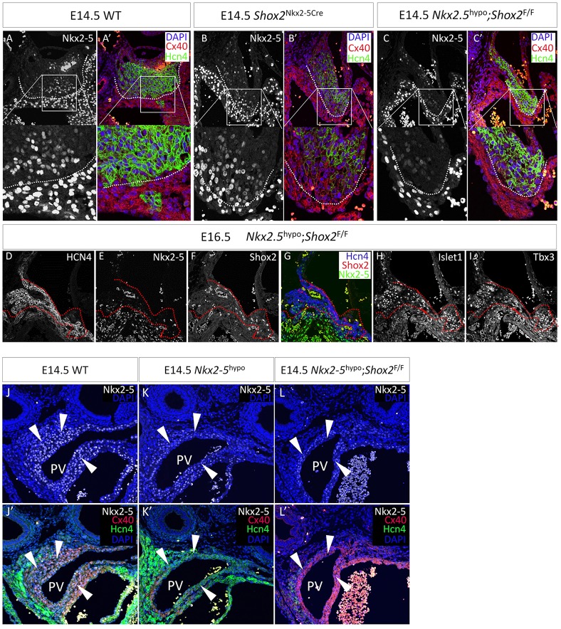
Shox2 antagonizes Nkx2-5 transcriptional output on the pacemaker program. (A-C′) A comparison of Hcn4 and Cx40 expression in the transition region of the Nkx2-5− SAN head and the Nkx2-5+ SA junction reveals a clear boundary (dotted lines) between the SAN cells and atrial cells in controls (A,A′), the conversion of Hcn4+/Cx40− cells to Hcn4−/Cx40+ cells in the Nkx2-5IRESCre/+;Shox2F/F (Shox2Nkx2-5Cre) SA junction (B,B′), and the rescue of this phenotype in Nkx2.5Cre/IRESCre;Shox2F/F (Nkx2-5hypo;Shox2F/F) mice (C,C′). (D-I) Expression of SAN markers, including Hcn4, Isl1 and Tbx3, is restored in the SA junction of Nkx2-5hypo;Shox2F/F mice. Dotted lines delineate the border between the SAN and atrial tissue that can be otherwise clearly observed in controls. (J-L′) Comparison of Hcn4 and Cx40 expression in the PV myocardium of control (J,J′), Nkx2-5hypo (K,K′) and Nkx2-5hypo;Shox2F/F (L,L′) mice. Arrowheads point to PV myocardium.

