
Fig. 7.
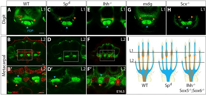
FDS tendon development is also modular and depends on muscle and cartilage. Transverse sections stained for MHC from (A,B) WT, (C,D) Spd and (E,F) Ihh−/− embryos at E16.5. Enlarged views of the boxed areas in B,D,F are shown in B′,D′,F′. Digit FDS tendons in (G) paralyzed mdg mutants and (H) Scx−/− mutants. (I) Schematic indicates digit and metacarpal levels and depicts the modular development of FDS tendons. Orange and blue arrowheads highlight FDS and FDP tendons, respectively. Scale bars: 50 μm.