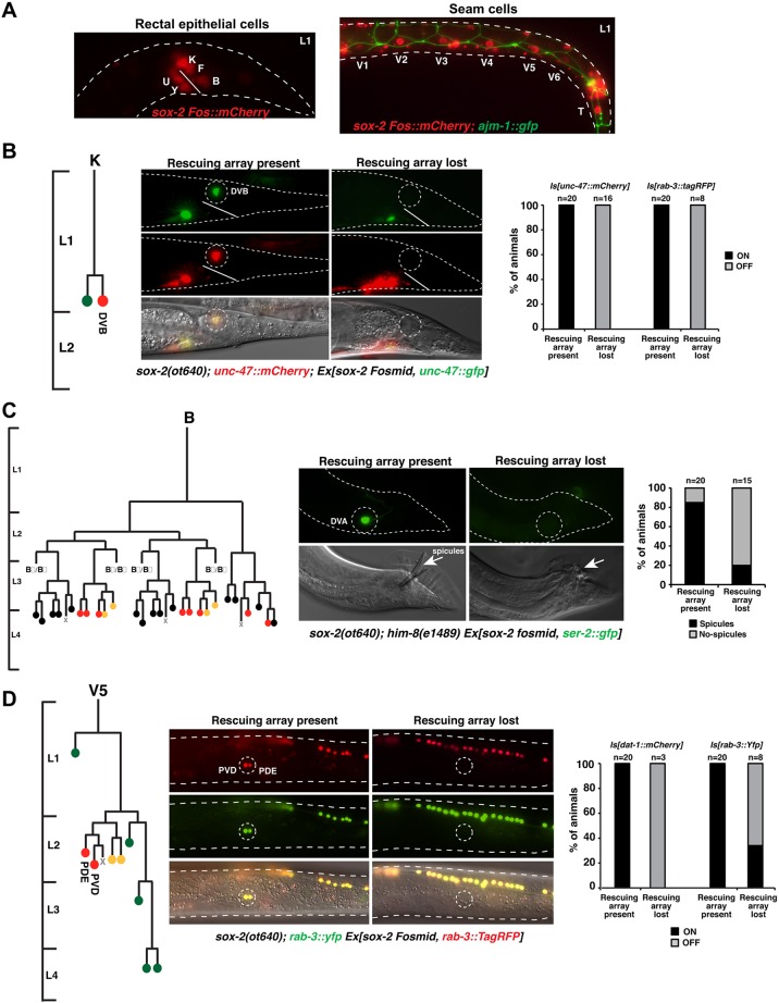
Fig. 3.
Postembryonic blast cells fail to generate certain lineages in sox-2 mutants. (A) Expression of sox-2 in rectal epithelial cells (left) and seam cells (right) at the L1 stage. (B) Mosaic analysis of sox-2(ot640) null mutants, showing loss of the DVB motor neuron. A scheme of the postembryonic K lineage is shown on the left. L1-lethal sox-2(ot640) animals were rescued with an extrachromosomal array (Ex) containing a fosmid with the sox-2 locus and the DVB reporter unc-47p::GFP in order to be able to follow the array in the K lineage. For phenotypic output, the expression of integrated (Is) fluorescent DVB (unc-47::mCherry) and pan-neuronal (rab-3::tagRFP) markers was analyzed. (C) Mosaic analysis of sox-2(ot640) null mutants, showing defective spicules in the male tail. Males were generated by crossing the sox-2(ot640) mutant into a him-8(e1489) mutant background. A scheme of the spicules-generating B lineage is shown on the left. L1-lethal sox-2(ot640) animals were rescued with an extrachromosomal array (Ex) containing a fosmid with the sox-2 locus and the DVA (sister of B) reporter ser-2p::GFP in order to infer the presence or absence of the rescuing array in the B lineage. For phenotypic output the presence of spicules in males was examined by DIC. (D) Mosaic analysis of sox-2(ot640) null mutants, showing loss of the PVD and PDE neurons. A scheme of the postembryonic V5 lineage is shown on the left. L1-lethal sox-2(ot640) animals were rescued with an extrachromosomal array (Ex) containing a fosmid with the sox-2 locus and the rab-3::TagRFP pan-neuronal reporter to assess the presence or absence of the rescuing array in the V5 lineage. For phenotypic output, expression of integrated (Is) fluorescent PVD and PDE markers (dat-1::mCherry and rab-3::yfp) was examined. Cell fate in lineage schemes: red, neurons; yellow, glial; green, hypodermal cells; black, proctodeum.

