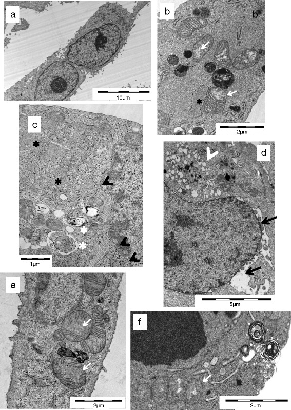
Fig. 7.

Electron microscopic images of cell death in thyroid carcinoma cells. Untreated SW1736 cells (a) with intact membranes, normal organelles, and morphology, (b) SW1736, (c) HTh7, (d) TPC1, (e) TPC1, and (f) BHT101 cells treated for 16 (c, d, e) or 24 h (b, f) with 0.1 μM GX15-070 as examples for swelling of mitochondria with loss of cristae (white arrows), blebbing of nuclear membrane (black arrows), extreme dilatation of rER with formation of reticular rER clusters (black asterisks), association of the nuclear membrane with membranes of dilatated rER black arrowhead), vacuoles probably originating from Golgi apparatuses (white arrowheads) as well as lamellar bodies and autophagosomes (white asterisks)
