Fig. 1.

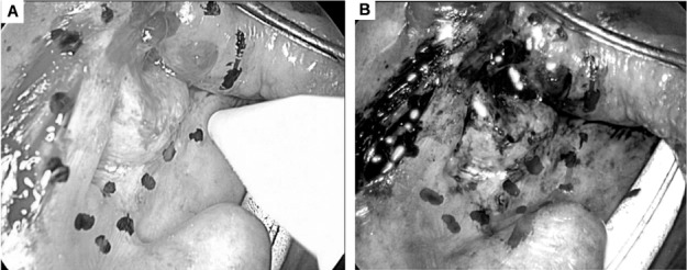
A) Marking the margins of a squamous cell carcinoma of the left tonsil under white light vision. B) Defining surgical margins with narrow-band imaging (red line showing further extension of the resection line).

A) Marking the margins of a squamous cell carcinoma of the left tonsil under white light vision. B) Defining surgical margins with narrow-band imaging (red line showing further extension of the resection line).