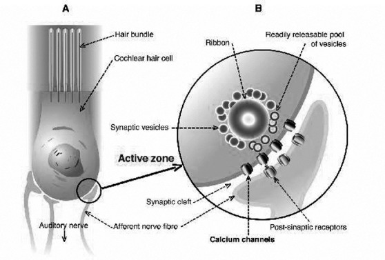
Fig. 2.

A) schematic representation of cochlear hair cell structure with baso-lateral wall cytoplasmic membrane (marked with circle) and afferent auditory nerve bottoms; and B) presynaptic "active zone" with vesicles and ribbon containing neurotransmitters with clustered voltage-gated calcium channel (VGCCs).
