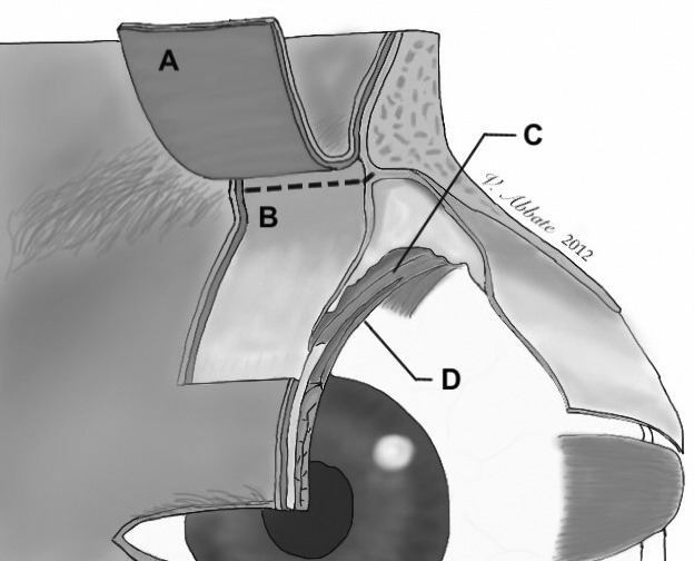
Fig. 1.

Left eye 3D cross-section view shows the plane of the transorbital approach and the supratarsal skin incision that is 10 mm above the tarsus, extending from the middle third of the upper eyelid to the lateral cantus. (A) Elevated muscolocutaneous flap, (B) dashed line shows the correct septal incision that reach the subperiosteal orbital roof plane, (C) levator palpebrae superioris, (D) superior tarsal muscle (Müller muscle).
