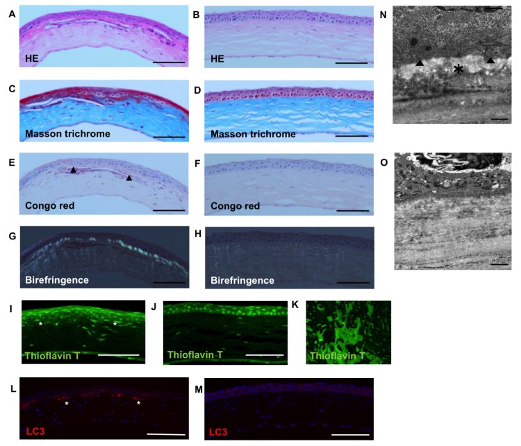
Fig 4. Histology of TGFBIR124H mice.
In homozygotes, HE staining did not show signs of inflammation in the cornea (A) as well as wild type mice (B), while Masson trichrome staining showed red deposits in the anterior cornea (C) in contrast to wild type mice (D). Congo red stating showed red deposits (arrow head) in the anterior cornea (E) and birefringence of the area was observed (G) in contrast to wild type mice (F, H). Subepithelial stroma was also stained with thioflavin T (*) in homozygotes (I) in contrast to wild type mice (J). Positive control: Human renal amyloidosis (K). Non-specific staining of cell nuclei is observed in both samples and positive control. Immunohistochemical examination with LC3 showed subepitheilaicl stromal staining (*) in homozygotes (L) in contrast to wild type mice (M). Examination with electronic microscopy revealed subepithelial deposits (*) in heterozygotes (N) in contrast to wild type mice (O). Autophagosomes (arrow head) were observed around the deposits. Scale bar = 100 um in A—J, 1 um in K, L.

