Fig. 4.

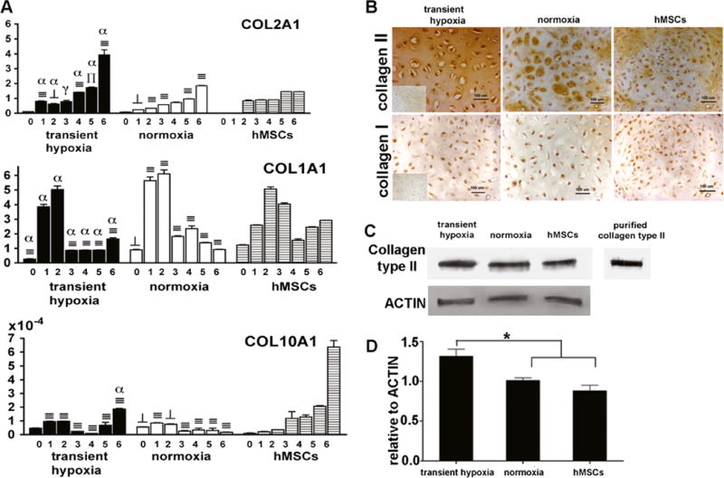
Extracellular matrix production in chondrogenic pellets. a Pellets derived from transient hypoxia EBs (Group 4), normoxia EBs (Group 2) and hMSCs were analyzed for gene expression of collagen type II, I and X. Gene expression levels were normalized with ACTB. Data show average ± SD of 4 samples. ⊥, ∏, ≡ denotes significant differences compared with hMSCs at the same time point, where p<0.05, 0.01 and 0.001, respectively. γ, β, α denote significant differences compared with normoxia, where p < 0.05, 0.01 and 0.001, respectively. b Immunohistochemistry showed localization of type II and type I collagen staining in 6 week pellets. Inserts show negative staining controls. c Bands specific for collagen type II protein were detected in pellet matrix using Western blot. Purified human collagen type II was used as positive control. d Relative intensities of collagen type II protein levels normalized to actin levels. Data show average ± SD (n=3).* denotes statistically significant differences (p<0.05)
