
Fig. 6.
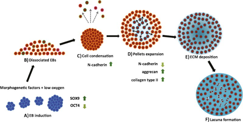
Model of chondrogenesis. a Embryoid bodies (EBs) were induced for 3 weeks by morphogenetic factors in chondrocyte-conditioned medium, under controlled oxygen tension (transient hypoxia regime). Induced EBs showed up-regulation of SOX9 and down-regulation of OCT4. b Chondrogenic progenitors mixed with non-chondrogenic progenitors are obtained after induction. c Chondrogenic progenitors expressing N-cadherin expression undergo cell condensation, while other cell populations are washed away with medium changes. d The spherical pellets increase in size as ECM production continues, resulting in increased intercellular spaces, starting at the middle of pellet and extending to the periphery. The progression of ECM synthesis occurs with a concomitant decrease in the expression of cell adhesion molecules. e ECM occupies entire intercellular spaces of chondrogenic pellets. The progenitor cells differentiate to chondrocytes. f After cell division, daughter cells located at the same area as parent cells secrete matrix to separate each other and form enclosed structures (lacunae)