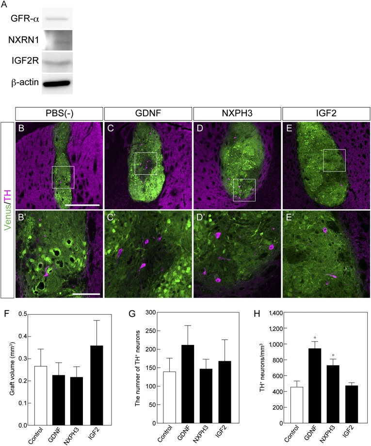
Figure 5.
Effect of soluble factors on induced pluripotent stem cell-derived (iPSC-derived) dopaminergic neurons in vivo. (A): Western blot analysis of receptors for GFR-α, α-NRXN and IGF2R. (B–E): Histological analysis of iPSC-derived grafts at 8 weeks with the indicated factor. Immunofluorescence images for venus (green) and TH (magenta). Scale bar = 50 μm. (F–H): Quantitative analysis of the graft volume (F), the total number of TH-positive (TH+) neurons (G), and the number of TH+ neurons per graft volume (H). Each value is given as the mean ± SEM (n = 5–6). ∗, p < .05 versus PBS(−) control group. Abbreviation: PBS(−), phosphate-buffered saline.

