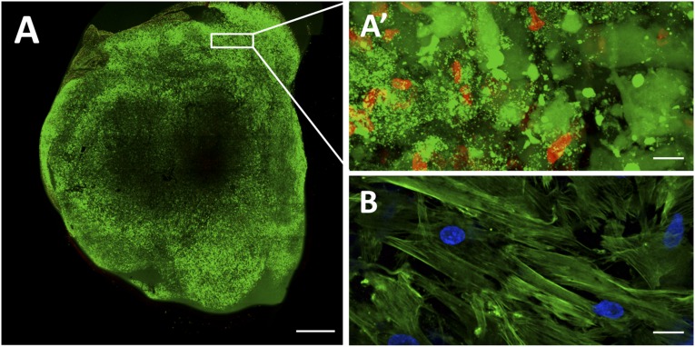
Figure 2.
Assessment of cell viability and differentiation status in the fibrin patch before in vivo implantation. (A): Representative image showing cell viability in a fibrin patch loaded with umbilical cord blood mesenchymal stem cells (UCBMSCs) and cultured for 24 hours in standard culture conditions. Viable cells are shown in green and dead cells in red, as performed using the Live/Dead viability/cytotoxicity kit (Invitrogen). (A′): Magnification of a selected peripheral area with a predominance of viable cells (green). (B): Fibrin patch immunostaining showing absence of CD31 expression (red) in UCBMSCs. Cell cytoplasm and nucleus were counterstained with Atto 488-phalloidin (green) and 4′,6-diamidino-2-phenylindole, respectively. Scale bars = 1 mm (A) and 20 μm (A′, B).

